
15 Jun learnECMO: a case of pulmonary HTN
53F initially presented to regional hospital with ~6/52 Hx of worsening SOBOE and type 1 respiratory failure. Initial investigations included a CTPA, which demonstrated no evidence of acute PE but features of both right heart strain and ?infective bronchiolitis.
The patient deteriorated on admission D4 with escalating oxygen requirements and underwent conventional medical retrieval to the regional tertiary referral centre. TTE demonstrated an RVSP ~100 mmHg with RV dilatation without hypertrophy. Within 24 hours she deteriorated further with a combination of progressive respiratory failure and obstructive shock requiring emergent intubation and then referral for ECMO retrieval for refractory hypoxaemia. She was stabilised on VV ECMO and eventually transferred to SVH for consideration of the best destination…
What is the differential diagnosis for this patient?
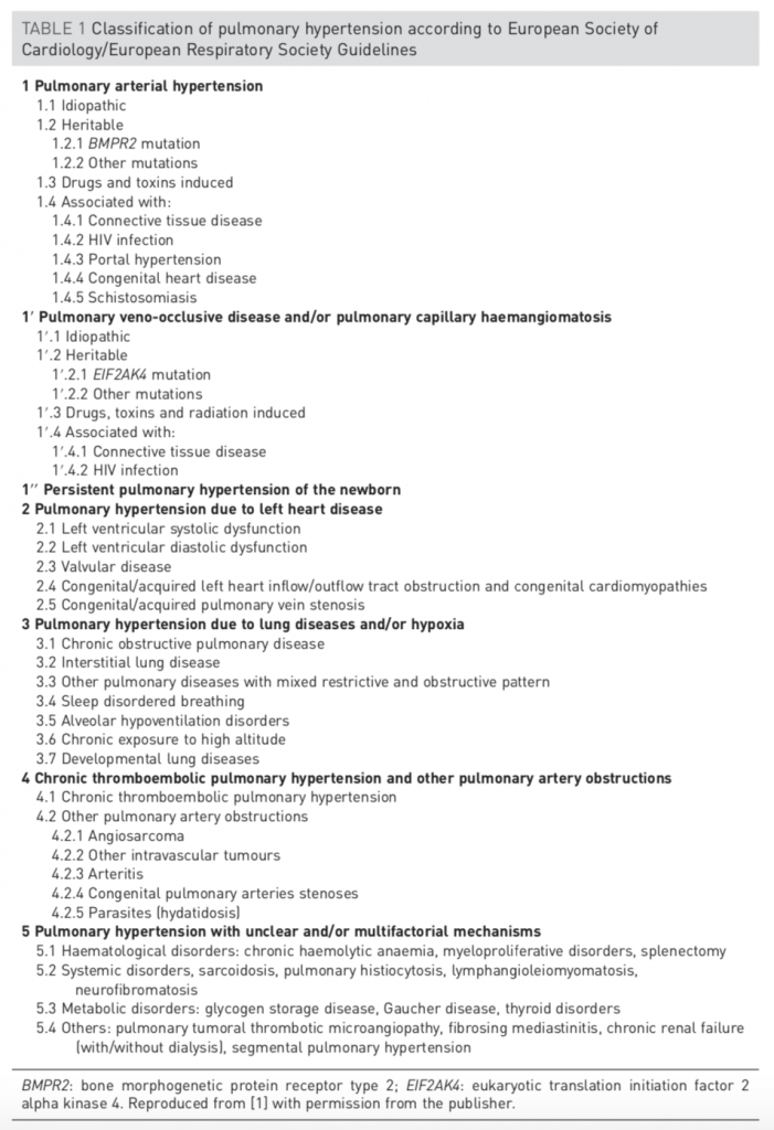
Differential diagnosis of pulmonary hypertension
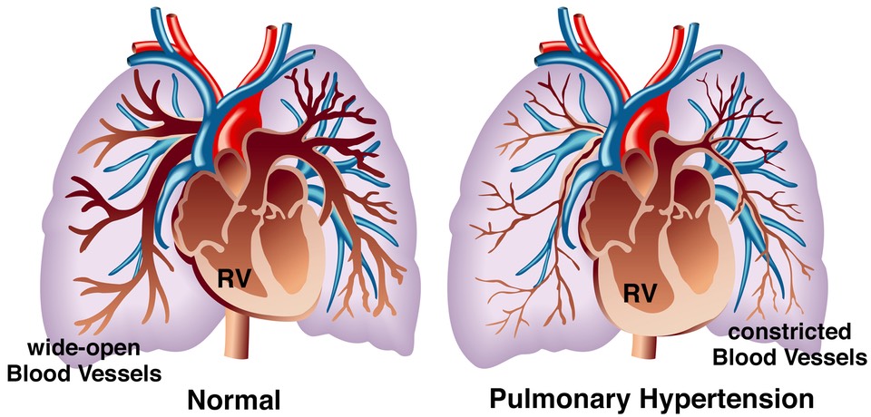
Pulmonary hypertension is a clinical syndrome characterised by mean pulmonary artery pressure ≥25 mmHg at rest on right heart catheter derived haemodynamic studies. It is sub-divided into 5 groups, of which, group 1 includes diverse pathophysiological entities that exhibit obliterative, small vessel pulmonary vasculopathy.
Observations & investigations
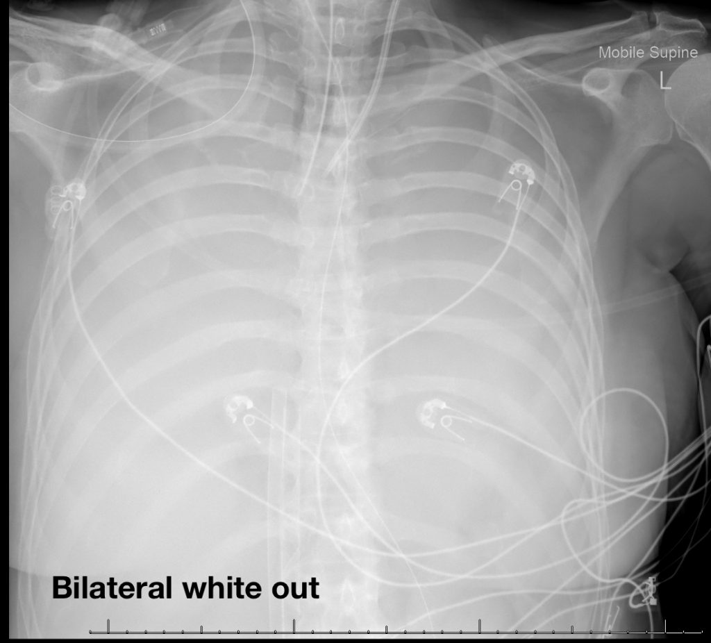
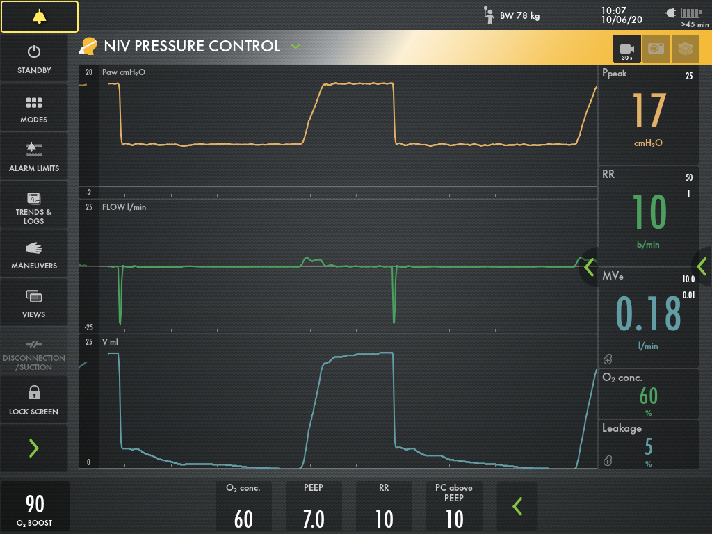
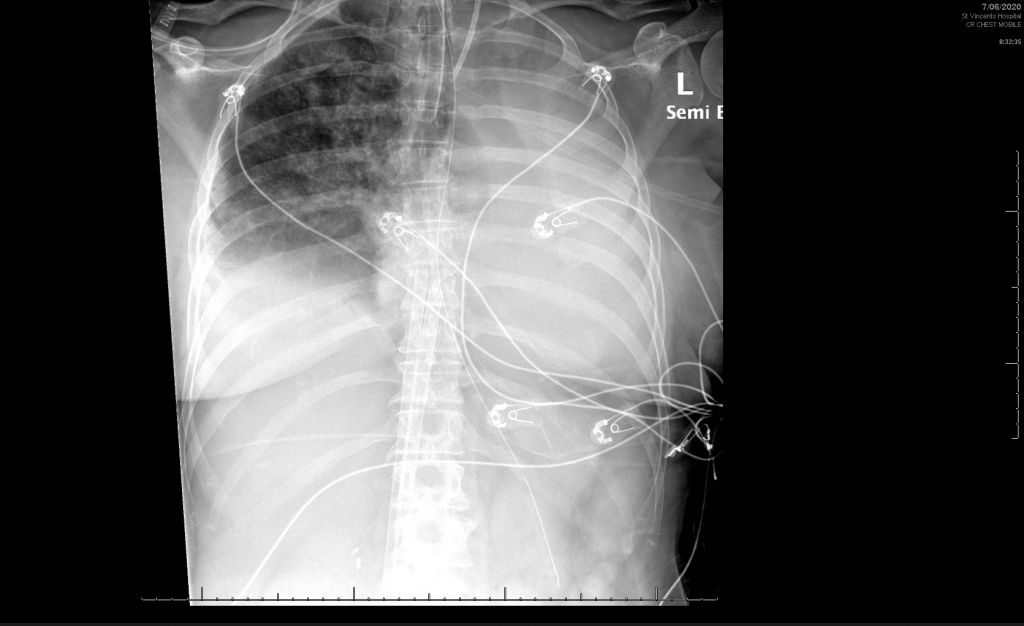
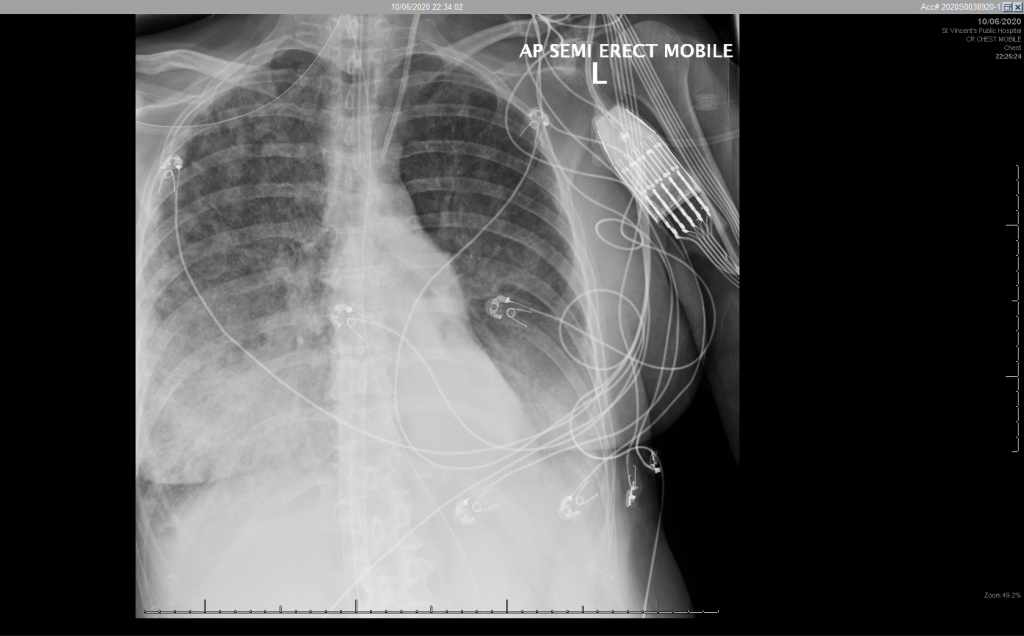
The patient had an initial working diagnosis of pulmonary capillary haemangiomatosis and thus had been referred for consideration for heart-lung transplant off VV ECMO. The patient experienced recurrent pulmonary haemorrhage with associated endobronchial obstruction and multi-lobar collapse, presenting as sudden reduction in tidal volumes and high airway pressures, requiring repeated therapeutic bronchoscopy.
Unfortunately, the patient died shortly after a one-way ECMO wean having been deemed not to be a transplant candidate for multiple reasons including previous infection with Burkholderia cepacia. The subsequent PM has not demonstrated a histological pattern consistent with pulmonary capillary haemangiomatosis, the full report is awaited.
Learning points
Pulmonary haemangiomatosis is a rare cause of pulmonary HTN
- PCH is a rare condition involving multiple angiomatous lesions of proliferating pulmonary capillaries
- Proliferating capillaries invade the pulmonary interstitium and alveolar septae leading to pulmonary vascular occlusion and eventually fatal pulmonary hypertension
- It is unclear if PCH is a distinct entity to pulmonary veno-occlusive disease (PVOD) [1]
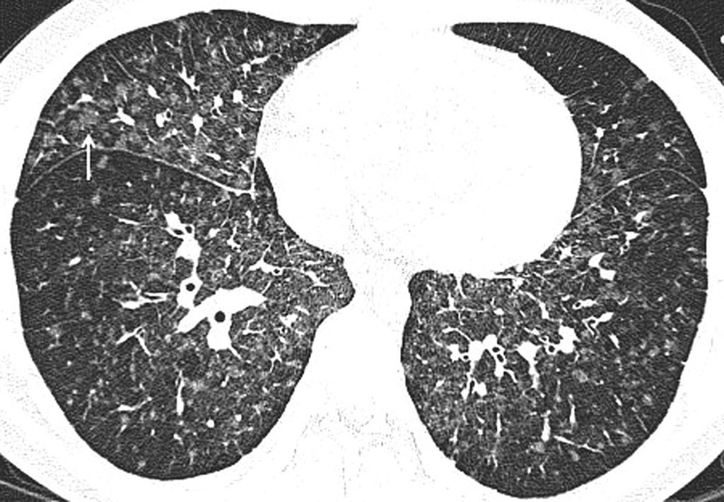
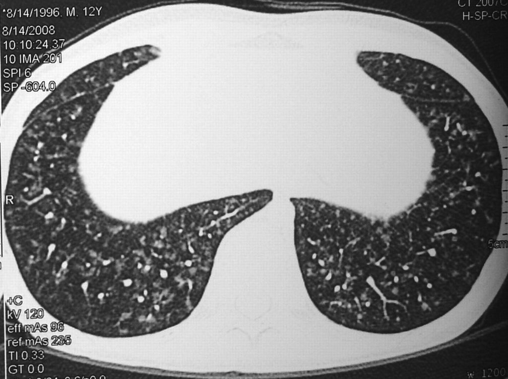
- PCH presents in a similar fashion to primary PAH but with distinct CT chest appearances including diffuse bilateral reticulonodular pattern with enlarged central pulmonary arteries [2]
- PVOD and PCH have been given the subgroup designation of group 1′ in order to highlight both similarities and important differences with PAH.
- In general, the clinical presentation and haemodynamic findings of PVOD/PCH and PAH are similar, but the clinical course of PVOD/PCH is usually more aggressive.
- Importantly, PVOD/PCH typically demonstrate a poor response to PAH therapy and the use of PAH drugs is associated with a potential risk of life-threatening pulmonary oedema [3]
- The natural history is one of inexorable decline, with heart-lung transplantation as the only therapeutic option to improve survival
1. Montani, D., et al., Pulmonary veno-occlusive disease. Eur Respir J, 2016. 47(5): p. 1518-34.
Burkholderia cepacia infection is a contraindication to lung transplant?
- Burkholderia (formerly Pseudomonas) cepacia complex consists of 18 distinct bacterial species that are obligate aerobic gram-negative rods
- BCC are ubiquitous environmental organisms with low human pathogenicity in immunocompetent hosts but can persist for extended periods in water and even disinfectants.
- Healthcare-related outbreaks have been linked to contaminated nebuliser solutions and IV medications and fluids.
- BCC emerged in the late 1970s as a CF respiratory pathogen associated with accelerated decline in lung function and reduced survival both pre- and post-lung transplantation. [1]
- BCC forms biofilms and demonstrates intrinsic resistance to a wide range of antimicrobial agents including: aminoglycosides, first and second generation cephalosporins, carboxypenicillins and polymyxin.
- The spectrum of associated clinical syndromes runs from colonisation to the Cepacia syndrome of necrotising pneumonia.
- BCC is subclassified as genomovars; species that are phenotypically similar and genotypically distinct. Genomovar III (B. cenocepacia) and genomovar II (B. multivorans) are the 2 most common isolates from CF patients. [2]
- Certain case series have demonstrated clinically significant post-transplant infection with the BCC strain that was isolated pre-transplant with excessive mortality associated with B. cenocepacia.
- Current infection with B. cenocepacia is variously considered a strong relative or absolute contraindication to lung transplantation. [3]
- Transplant candidacy must always consider the broad scope of patient-specific factors that influence the probability of early mortality. [4]
-
- Corey, M. and V. Farewell, Determinants of mortality from cystic fibrosis in Canada, 1970-1989. Am J Epidemiol, 1996. 143(10): p. 1007-17.
- Aris, R.M., et al., Lung transplantation for cystic fibrosis patients with Burkholderia cepacia complex. Survival linked to genomovar type. Am J Respir Crit Care Med, 2001. 164(11): p. 2102-6.
- De Soyza, A., et al., Lung transplantation for patients with cystic fibrosis and Burkholderia cepacia complex infection: a single-center experience. J Heart Lung Transplant, 2010. 29(12): p. 1395-404.
- Weill, D., et al., A consensus document for the selection of lung transplant candidates: 2014–an update from the Pulmonary Transplantation Council of the International Society for Heart and Lung Transplantation. J Heart Lung Transplant, 2015. 34(1): p. 1-15.
VV ECMO for pulmonary HTN?
- The selection of the appropriate ECMO configuration for a patient with concomitant severe respiratory and cardiovascular failure is challenging.
- Detailed assessment of the mechanisms of shock incorporating echocardiography is essential and should be contextualised to the patient’s presentation and pre-morbid cardiac function.
- Severe ARDS may be associated with acute right ventricular failure or acute cor pulmonale (ACP). [1]
- In SRF with ACP there is evidence that VV ECMO initiation offloads the associated acute pulmonary hypertension and hence RV afterload. This is likely multifactorial related to normalised gas tensions, increased pH and altered heart-lung interaction associated with ultra-protective ventilation settings. [2, 3]
- Primary PAH associated with RV failure and refractory cardiogenic shock without severe hypoxaeamia is likely best managed with VA ECMO to support the circulation as a bridge to RV recovery or transplantation. [4]
1. Mekontso Dessap, A., et al., Acute cor pulmonale during protective ventilation for acute respiratory distress syndrome: prevalence, predictors, and clinical impact. Intensive Care Med, 2016. 42(5): p. 862-870.
2. Reis Miranda, D., et al., Right ventricular unloading after initiation of venovenous extracorporeal membrane oxygenation. Am J Respir Crit Care Med, 2015. 191(3): p. 346-8.
3. Grant, C., Jr., et al., ECMO and Right Ventricular Failure: Review of the Literature. J Intensive Care Med, 2020: p. 885066619900503.
4. Machuca, T.N. and M.d. Perrot, Mechanical Support for the Failing Right Ventricle in Patients With Precapillary Pulmonary Hypertension. Circulation, 2015. 132(6): p. 526-536.